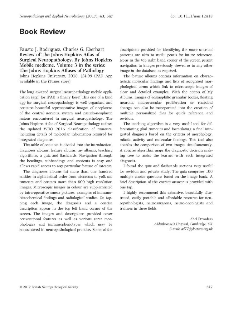
The following review recently appeared in the journal Neuropathology and Applied Neurobiology:
The long awaited surgical neuropathology mobile application (app) for iPAD is finally here! This one of a kind app for surgical neuropathology is well organized and contains beautiful representative images of neoplasms of the central nervous system and pseudo-neoplastic lesions encountered in surgical neuropathology. The Johns Hopkins Atlas of Surgical Neuropathology utilizes the updated WHO 2016 classification of tumours, including details of molecular information required for integrated diagnoses.
The table of contents is divided into the introduction, diagnoses albums, feature albums, my albums, teaching algorithms, a quiz and flashcards. Navigation through the headings, subheadings and contents is easy and allows rapid access to any particular feature of interest.
The diagnoses albums list more than one hundred entities in alphabetical order from abscesses to yolk sac tumours and contain more than 800 high resolution images. Microscopic images in colour are supplemented by intra-operative smear pictures, examples of immunohistochemical findings and radiological studies. On tapping each image, the diagnosis and a concise description appear in the top left hand corner of the screen. The images and descriptions provided cover conventional features as well as various rarer morphologies and immunophenotypes which may be encountered in neuropathological practice. Some of the descriptions provided for identifying the more unusual patterns are akin to useful pearls for future reference. Icons in the top right hand corner of the screen permit navigation to images previously viewed or to any other image in the database as required.
The feature albums contain information on characteristic molecular findings and lists of recognized morphological terms which link to microscopic images of clear and detailed examples. With the option of My Albums, images of eosinophilic granular bodies, floating neurons, microvascular proliferation or rhabdoid change can also be incorporated into the creation of multiple personalized files for quick reference and revision.
The teaching algorithm is a very useful tool for differentiating glial tumours and formulating a final integrated diagnosis based on the criteria of morphology, mitotic activity and molecular findings. This tool also enables the comparison of two images simultaneously. A concise algorithm maps the diagnostic decision making tree to assist the learner with each integrated diagnosis.
I found the quiz and flashcards sections very useful for revision and private study. The quiz comprises 100 multiple choice questions based on the image bank. A brief description of the correct answer is provided with one tap.
I highly recommend this extensive, beautifully illustrated, easily portable and affordable resource for neuropathologists, neurosurgeons, neuro-oncologists and trainees in these fields.
Abel Devadass
Addenbrooke’s Hospital, Cambridge, UK
E-mail: [email protected]
