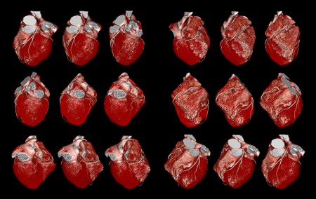
Before Friday, May 31, 2013, you need to go to the Turner Concourse located on Rutland and Monument Streets to see the magnificent exhibition entitled, Hidden Beauty: Exploring the Aesthetics of Medical Science!
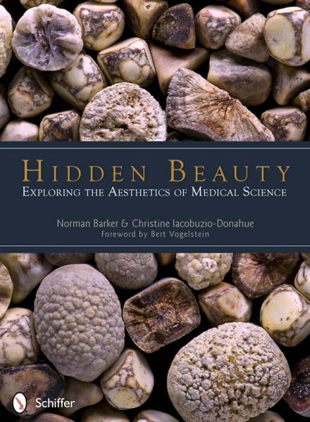
This exhibit features 50 amazing images and corresponding descriptions from the newly published book by Pathology faculty, Norman Barker and Dr. Christine Iacobuzio-Donohue. Over 60 medical professionals, including many from Johns Hopkins, contributed to this book with the same title, Hidden Beauty: Exploring the Aesthetics of Medical Science. The book cover is unusual because on close inspection you realize this beautiful image is actually a mound of gallstones.
Each of the exhibit’s works is fascinating, very wonderful, and engaging to the viewer. Many of the images are visually stunning patterns of different diseases that affect mankind. One leaves the exhibit with a great appreciation for biology in general, the wonder of the human body, for medical scientists, the study of the particular type of science they have dedicated their lives to, the technology or talent that enables us to see the image, and last but certainly not least, the artistry behind the image.
The description provided for each picture helps the viewer to better understand and appreciate the subject matter, the science, why it was chosen, and how it was created. Because it is impossible to fully describe these beautiful images and because of space constraints, I will provide an brief overview
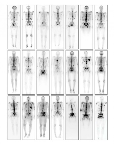
The spectrum of these extraordinary medical science images is far-ranging — from an engaging electron microscopic images of telomerase, to a colorful photo of an actual research bench with its descriptive analogy of a work bench being similar to the kitchen in a busy restaurant, to the very sad and poignant bone scan of patients with metastatic prostate cancer. A “nude” mouse is featured in one selection because these naturally hairless creatures are invaluable to certain types of research because they do not mount a rejection response to human tumor grafts.
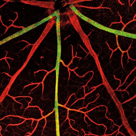
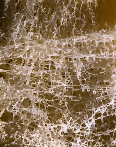
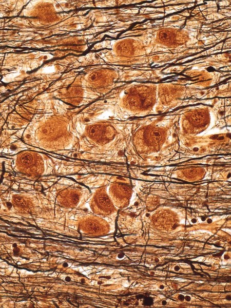
The kidney images were taken with a scanning electron microscope and a transmission electron microscope that can magnify 400x to 10,000X and depict the most detailed imagery available. The human brain image is fascinating — a 400X magnification of neurons, dendrites and axons. The nine images of the human eye, one normal and eight diseased are exceptional. Other presentations include extraordinary images of gallstones (featured on the book cover), HIV, osteoporosis, DNA sequencing, TB bacterium, Pap smear, tissue microarray, the placenta, salmonella, Max Broedel’s illustration of the inner ear, much more.
Download the poster here.
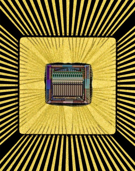
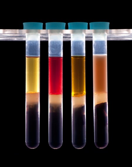
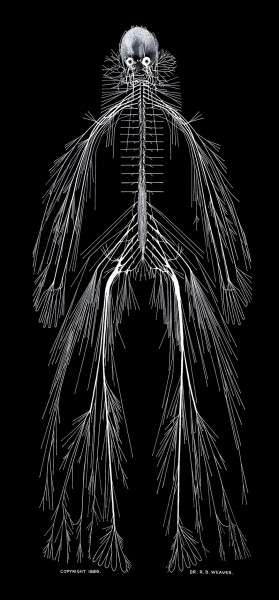
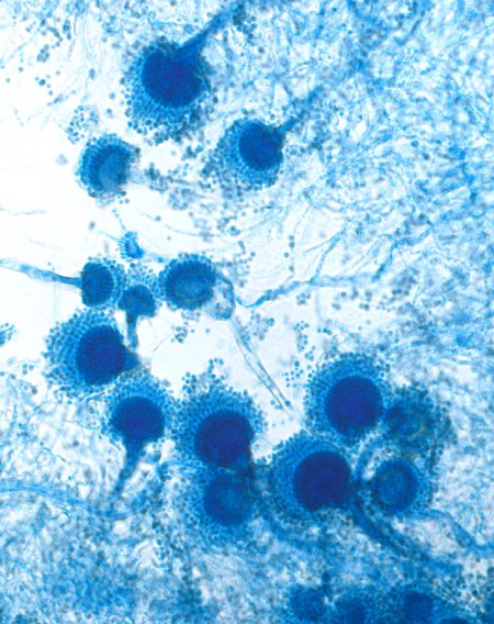
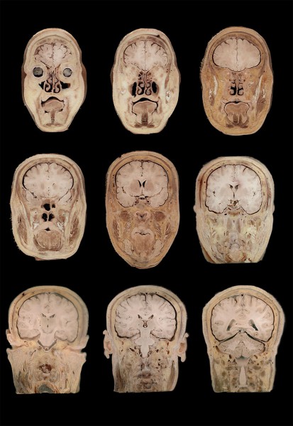
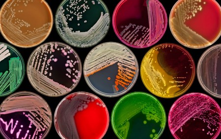
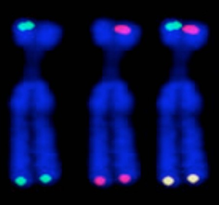
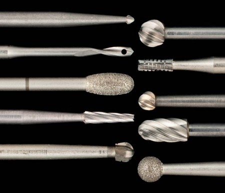
Look below to see some of the imagery from this exhibit (click for larger) which runs until Friday, May 31. Please do not miss it!
Renata Karlos
Staff Assistant
Department of Pathology
