 In almost every lab across the country, efforts to improve turnaround times, provide better service to patients and maintain a safe environment for staff are at the forefront. We are no different, if anything we have higher expectations placed upon us as the premier hospital in the nation. It is because of this fact that an effort to change “how we’ve always done it” has come with mixed reviews.
Routinely, the processing of a specimen in Surgical Pathology begins in accessioning, then moves to grossing for sectioning and is placed into fixative, normally 10% Neutral Buffered Formalin. The tissue cassettes are then picked up by a Histo tech at the end of the day, loaded onto a standard tissue processor on a 12 hour overnight processing schedule. The goal of processing is to remove all of the water from the tissue through a series of graded alcohols and xylenes; and then infiltrate the tissue with paraffin wax. This will allow the tissue to then be embedded, sectioned and placed on a slide for staining and pathologists’ review. This process has not changed in almost 100 years, this is histology.
Recently, as in the past 30 years, microwave technology has been explored as an alternative to routine processing. Microwaves (electromagnetic waves) are short pulses of energy that allow the solution that the tissue cassettes are submersed in to be heated evenly and are able to maintain that temperature over time. The overall benefit from this heat element alone is that the fixation and dehydration of the tissue occurs in about one third of the time. In essence, a routine specimen such as a radical prostatectomy, grossed at a thickness of 3mm, can be processed by our microwave processor in about 3 hours 45 minutes in comparison to a standard 12 hour processing schedule.
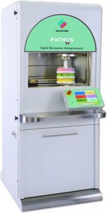
 The instrument that is used in the histology lab is the Pathos, manufactured by Milestone Medical Inc. Some of the features include separate retorts for the microwave chamber and the paraffin chamber. This allows us to run cycles back to back without the need to run a cleaning cycle after each run. There are multiple preset programs for tissue thicknesses from 1 mm to 5 mm, and the ability to customize our own processing cycles. These processing times run anywhere from 1 hour to 5 hours depending upon the thickness of the tissue. We utilize an isopropyl/ethanol alcohol blend to eliminate the need to use xylene. This reduces hazardous chemical exposure by the staff and reduces the overall amount of hazardous waste produced by the lab. Pathos, has a solution manager built in to the software. Based on the number of cassettes that are processed, the software calculates the usage and lifespan of the solutions. This allows us to monitor the exchanges and consumption of reagents much more accurately. The ease of the software makes the instrument very user friendly and the feedback from the staff has been very positive.
We have faced many challenges in our validation process. Through trial and error, our tissue collection has improved dramatically. A coordination of efforts between the histology and grossing staff has allowed us to grow our tissue bank very rapidly in recent weeks. In a department this large, each service has its own set of requirements they would like to evaluate before signing off on the change in processing. This has created some delays in getting certain tissue types approved. While trying to advance the technology and improve the turnaround times of the specimens, we also want to show that microwave processing will give comparable results to the current method in a much shorter time frame.
One specific area that has been very sensitive to validate are the breast specimens. We have shown that the processing can be performed on large mastectomy samples in less than 5 hours and quality sections are being produced. The main Achilles’ heel with this tissue type is validating a large enough sampling of Her2Neu- 2+ positive cases. The additional breast immuno stains (ER/PR, Ki67, GCDFP and Mammoglobin) have resulted in comparable stain quality and clarity.
All in all, we have found the Pathos microwave processor to be an asset to the workflow and help us to distribute the workload more effectively. I would like to thank the pathologists’ who were very eager and excited to get on the microwave bandwagon to help us get this project started and appreciate their continued support.
You can learn more about the Pathos workflow on the milestone medical web site.
Deborah Duckworth, HTL (ASCP)
Histology Supervisor