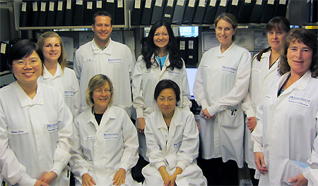
Flow Cytometry lab Members
A patient comes into the ER with fever, bruising, fatigue. Results from Hematology show a high white blood cell count, and blasts seen on the peripheral blood differential. The patient’s family gets the painful news that this appears to be Leukemia and the sooner treatment begins, the better. The patient is admitted to Weinberg. Everything is happening so fast. The correct treatment is essential in overcoming this disease and getting the patient to a possible recovery, but wait! Who can help in determining what type of disease we are dealing with so the Oncologists can determine the best treatment?
Flow Cytometry has become a very important technique used to assist in the diagnosis of Leukemia and Lymphomas. The technique itself can be used for many applications in the clinical and research fields. The Johns Hopkins Clinical Flow Cytometry Lab under the direction of Dr. Michael Borowitz, a world renowned expert in the field, provides testing for Leukemia, Lymphoma, PNH, T cell subsets, Quantitative CD34, and Minimal Residual Disease of Leukemia or Lymphoma.

Click to view larger *
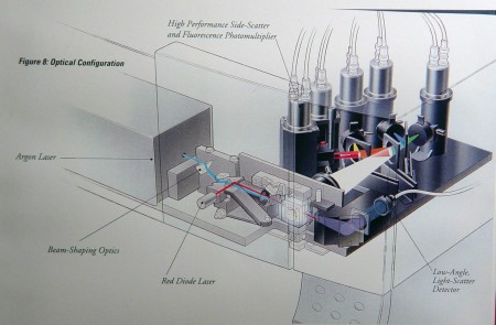
Using a technique where antibodies attached to a fluorescent dye are used to check for specific antigens on the surface of cells, one can determine or at least narrow down the identity of a cell. If the antigen being tested for is present, the laser in the Flow Cytometer excites the fluorescent dye and it emits at a certain wavelength. Depending on how many different dyes can be detected by the flow cytometer enables you to use more than one antibody at a time. These patterns are compared to normal patterns to determine the significance of the results.
The test takes approximately three hours and consists of staining the cells, acquiring the cells on a flow cytometer, and then having a skilled technologist analyze the results that have been saved to a computer file. Flow Leads, Fellows, and Attendings review and interpret these results to provide the patients and their physicians with the most accurate disease assessment. In most instances, the patient can be properly diagnosed within a matter of hours and the correct treatment can get underway.
![]()
Laser Light Euroscicon *
Besides blood and bone marrow, Flow staff are trained in preparation of fine needle aspirates, lymph nodes, tissue, CSF, other fluids, and even pieces from organs. All cells must be viable so samples cannot be fixed and should arrive to the Flow lab right away. One of the challenges that faces Flow is determining normal populations from abnormal populations, especially when looking for minimal residual disease after the patient has been treated. Progress and experience in this area has given the Johns Hopkins Clinical Flow Lab an excellent reputation.
If you have any questions or would like to learn more, stop by and see us sometime. We are located in Weinberg Room 2300.
Kathleen Cowan, MT(ASCP)
Technical Specialist
Flow Cytometry
Division of Hematology
* photo credits to bdbiosciences.com and eurosciconnews.com
Hi Kathy and Team,
I have just seen this picture of the flow team on the pathology website, and wanted to say hello!
I am still in Zimbabwe, but getting ready to move to South Africa now.
Please say hello to the rest of the team.
Yaw Agyei.
Yaw, wonderful to hear from you and happy to know you keep up with the Pathology website. Best wishes on your move.
From Kathy and the Flow Lab
Hi Yaw. We miss you. It is great to hear that you are doing well.
Jen Taylor (Hurley)
Yaw!!! We miss you here!!!