The time I spent viewing the current exhibition, “Celebrating 100 Years Teaching Excellence in Medical Illustration,” has been one of the highlights in my 20 years of working for this renowned Institution. I urge you not to miss the opportunity to view the rich and varied artwork of this extraordinary Centennial Exhibit. It will fill you with awe and appreciation for the great contributions that medical illustrators have given to the medical and scientific profession. The exhibition will make you marvel at the amazing intricacy of the human body, the enormous talent of medical illustrators, and the trajectory the profession has taken over the past 100 years to produce art for medical science. The collection includes an array of subjects — anatomy, pathologic specimens, surgical techniques, textbook illustrations, magazine covers, and more — created with pen and ink, carbon dust, watercolor, photography, and digitized media.
Included in this Blog post are reproductions of some of the works graciously provided by the Johns Hopkins University Department of Art as Applied to Medicine. Artists include Max Brödel (1870-1941), the founder of the Department, current and former faculty, graduates, and students. All art is copyright-owned by the individual artists, and no art may be reproduced without permission.
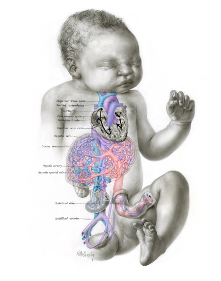
Ranice W. Crosby: Cardiovascular System of Fetus in Utero
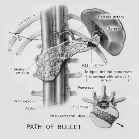
Duncan Winter: Path of Bullet: McKinley’s Assassination
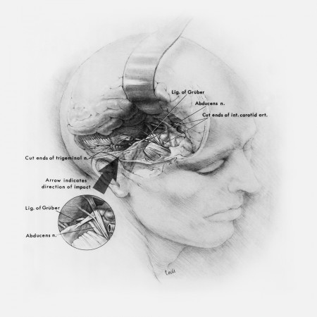
Al Teoli: Surgical Exp. to the Abducens Nerve
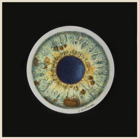
Sharon Weilbacher: An Unusual Iris Melanoma
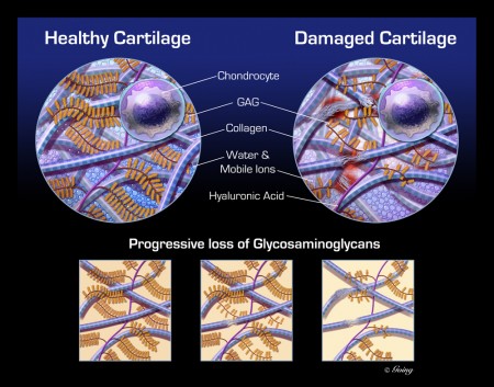
Ellen Going: Osteoarthritis Change in Articular Cartilage
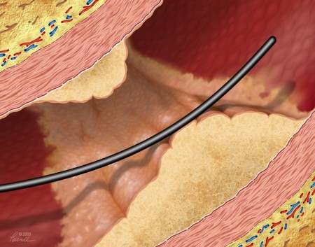
David R. Purnell: Atherosclerotic Plaque in an Artery
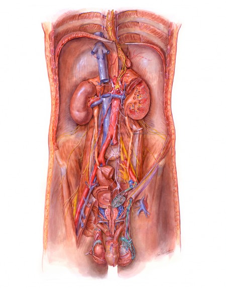
Leon Schlossberg: Genitourinary Tract – Vessels and Nerves
Some readers may not be aware that the Department of Art as Applied to Medicine was founded in 1911 by Max Brödel, a German émigré. Brödel arrived in Baltimore in 1894 and immediately began to work for Dr. Howard A. Kelly in the Gynecological Department of Johns Hopkins Hospital. Several months later Brödel began to plan his medical art curriculum. In 1911 Brödel founded the Department of Art as Applied to Medicine, the first academic department of medical illustration of its kind in the world. Henry Walters, philanthropist, art collector and founder of the Walters Art Gallery, endowed the Department of Art as Applied to Medicine, establishing its permanence.
Max Brödel is considered by most medical illustrators to be the father of the profession. Clearly it was not just his artistic talent that made him so great, but his great intellect, his deep-driven curiosity, his hard work, and his love of learning. Below is an interesting link about Brödel, written by his friend and colleague Dr. Thomas S. Cullen of Johns Hopkins: http://www.ncbi.nlm.nih.gov/pmc/articles/PMC200894/pdf/mlab00259-0010.pdf.
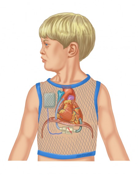
Jon Coulter: Pedia Flow Adolescent Heart Pump
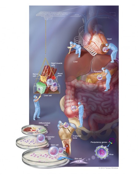
Therese Winslow: The Other Stem Cells
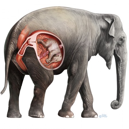
Dr. Levant Efe: Pregnant Elephant
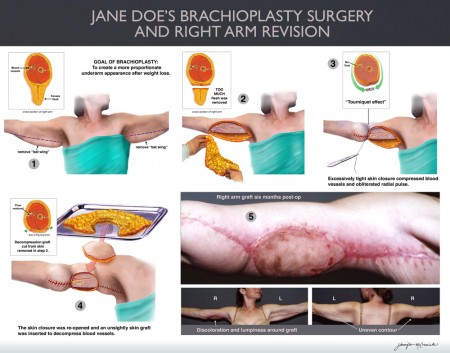
Jennifer (Graeber) McCormick: Brachioplasty and Right Arm Revision
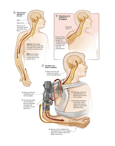
Michael Linkinhoker: Neural Arm Prosthetic
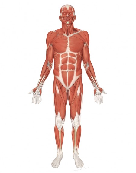
John Gibb: Principle Superficial Skeletal Muscles
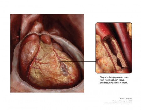
Ann G. Canapary: Myocardial Infarction
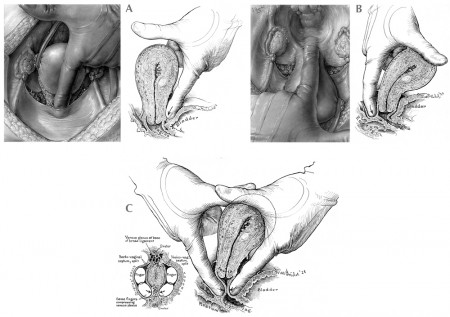
Timothy Phelps, the Assistant Director and Associate Professor of the Department of Art as Applied to Medicine, gave me a tour of the Exhibit which starts with medical illustrations from the early 1900s, when the artists often used carbon dust as their medium. I had never heard of carbon dust. Tim told me that the carbon dust technique was mastered by Max Brödel. Carbon dust is made by shaving the carbon from a pencil to create a dust. The dust is then applied with a paintbrush onto calcium-coated paper. This drawing method renders images of exquisite visual detail still and is still used by medical and scientific illustrators 100 years later. Below is an example of Brödel’s work with pen and ink and carbon dust:
Max Brödel: Uterus
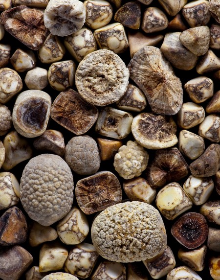
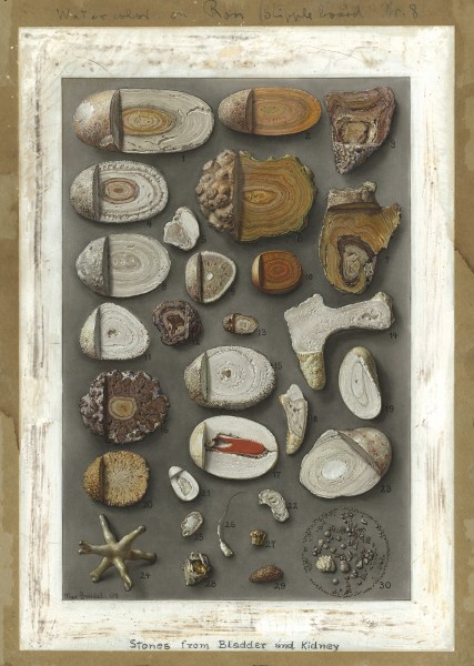
I also want to mention that Pathology faculty, Norman Barker, Associate Professor of Pathology and Director of Pathology Photography, has on display a very interesting, almost artsy photograph of gallstones. When I asked Norm why he chose this particular piece from his huge collection of prints, he chuckled and replied that his photograph was in homage to Max Brödel who was well known for his illustration of kidney stones, first published in 1914. (Who would have known that gallstones and kidney stones could be so beautiful!)
Norm has a Joint Appointment in the Department of Art as Applied to Medicine as an Instructor of Photography and digital media, and told me that today’s students study computer animation and modeling as part of their two-year program. The curriculum evolves with the times.
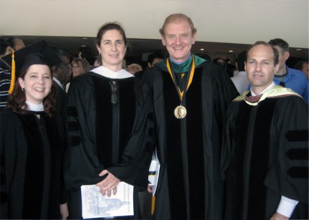
Also, in May of this year at the Johns Hopkins University Medical School graduation, Pathology Professor Ralph Hruban received the highest award from the Department of Art as Applied to Medicine, the Ranice W. Crosby Distinguished Achievement Award, for scholarly contributions to the advancement of art as applied to the medical sciences. Dr. Hruban was saluted for his investigative spirit to explore novel uses of new and innovative visual technologies to communicate medicine. The Art as Applied to Medicine Department commended Dr. Hruban not only for his acknowledgement of the visual representation of scientific information by the artist as an essential component of medical education, but for actively partnering with the illustrators to create educational images.
Left to right: Department of Art as Applied to Medicine faculty Assistant Professor Jennifer Fairman and Associate Professor Cory Sandone, Award winner Professor Ralph Hruban of Pathology, and Associate Professor David Rini of Art as Applied to Medicine
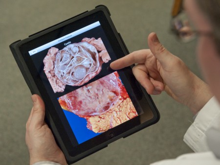
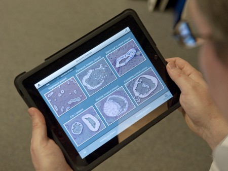
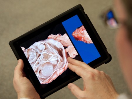
Over the past year Dr. Hruban worked with Art as Applied to Medicine graduate student, Bona Kim (’11), to develop a novel iPad application to teach pancreatic pathology. Bona was awarded both a Vesalius Trust Grant and the Inez Demonet Scholarship, the latter being the top national award for art as applied to medicine students, for this work. See pictures of their collaborative product:
Congratulations, Dr. Hruban and Bona!
More pictures:
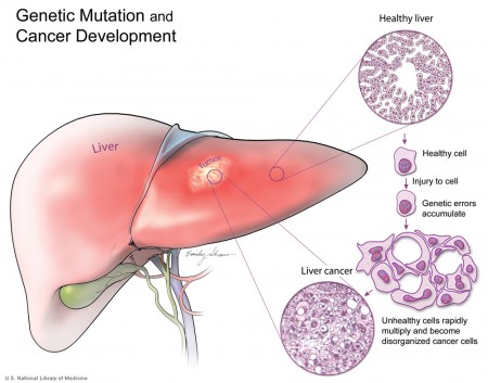
Emily Shaw: Genetic Mutation and Cancer Development
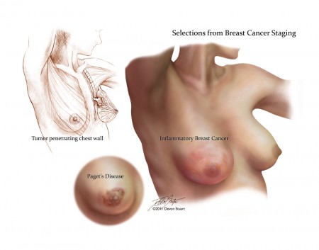
Devon Stuart: Selections for Breast Cancer Staging
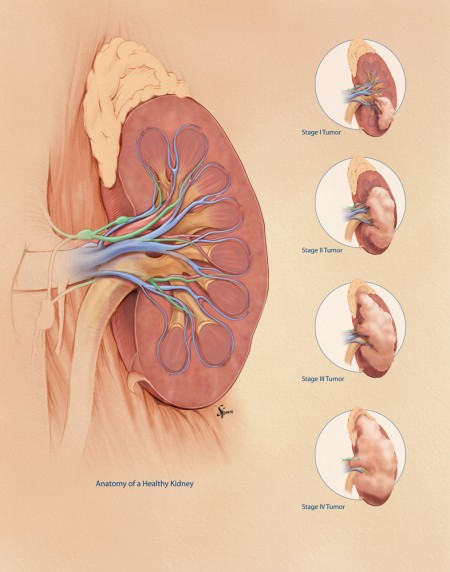
Satyen Tripathi: Understanding Renal Carcinoma
Jenny Wang: Debris in our Oceans
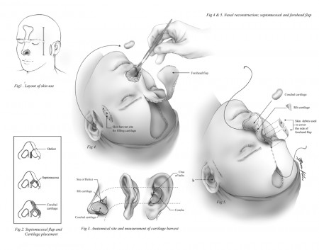
Bona Kim: Nasal Reconstruction with Forehead Flap
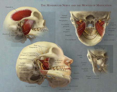
Elyssa L. Siegel: Mandibular Nerve and Muscles of Mastication
Dacia Balch, the Admissions Coordinator for Art as Applied to Medicine, told me that admission to the prestigious program is highly competitive — only 8-12% of all candidates are accepted. Dacia described the current exhibit as “spectacular, unique, one of a kind.” The Department is working on a Centennial Exhibition catalogue to be released in the very near future.
Finally, my readers, do take the time walk over to the Turner Concourse to view this marvelous exhibit, Celebrating 100 Years Teaching Excellence in Medical Illustration. You will undoubtedly be amazed and awestruck by the level of artistic work on display. You will come away with a great admiration and appreciation for the work of Max Brödel and his legacy of medical illustrators. The exhibit will run until November 15, 2011 in the Turner Concourse. Make plans to attend!
Special thanks to Tim Phelps of the Department of Art as Applied to Medicine and Norm Barker of Pathology Photography and Graphic Arts for their time and assistance with this Blog post.
Renata Karlos
Staff Assistant
Department of Pathology
Ranice W. Crosby: Cardiovascular System of Fetus in Utero

Great write up! I love the photos – you really went all out on this one.
Thank you for sharing and your interest.